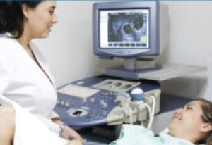

Why is an Ultrasound Done?
Ultrasonographic assessment is an extremely important part of any fertility assessment because it identifies any problems or irregularities which may affect conception, providing your Specialist with information to facilitate treatment planning. The transvaginal ultrasound picture depicts accurately the pelvic anatomy of the scanned area safely, quickly and reproducibly.
USG assessment of the endometrium is an important investigative tool in the assessment of receptivity of endometrium.
Congenital uterine abnormalities may have an important role in subinfertility, implantation failure and outcome of pregnancy
Endometrial volume is a useful criterion in predicting embryo implantation success and pregnancy rate in patients undergoing in-vitro-fertilization (IVF).
Why use 3D/4D Technology?
3D/4D scanning technology is the most advanced technique in ultrasound. In pre-conception imaging, 3D ultrasound is particularly useful for identifying abnormalities in the uterus, it helps in the evaluation of the uterine cavity for the presence of endometrial polyps (benign growths of the lining of the uterus), uterine fibroids (benign tumours of the muscle of the uterus) or other conditions such as endometriosis.
3D and 4D scanning provides:
- More detailed imaging
- The most effective identification of issues which might affect fertility or the possible causes of recurrent pregnancy loss
- Information which will assist with prediction of your response to the drugs used to stimulate your ovaries
- Planning of optimum treatment