
01
HSG X-Ray
Uterus and Fallopian Tubes

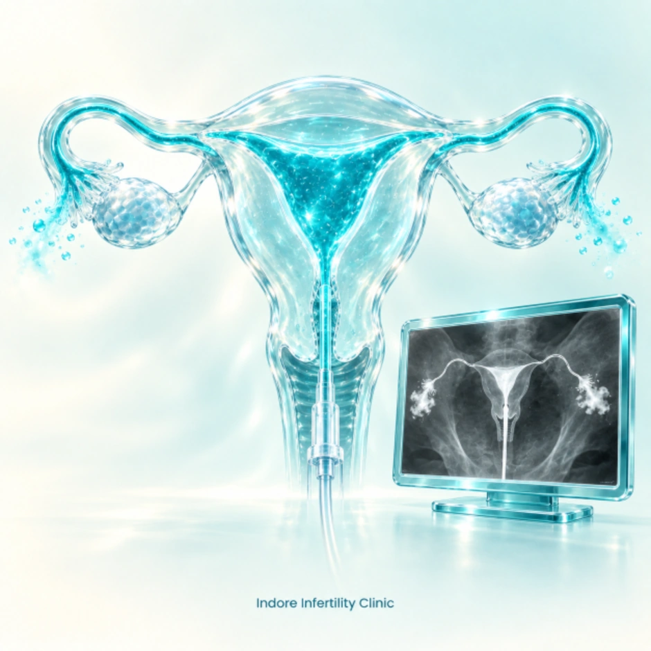
Hysterosalpingography, commonly called HSG, is an important fertility investigation used to examine the uterus and determine whether the fallopian tubes are open. It is an X-ray based procedure in which a special radiographic contrast dye is introduced into the uterus through the vagina and cervix. As the contrast travels through the uterine cavity and fallopian tubes, a series of X-ray images are taken to assess the shape and outline of the uterus and to see whether the tubes allow the dye to pass through normally. HSG is commonly recommended as part of female fertility evaluation because blocked or damaged fallopian tubes can interfere with the normal movement of the egg and sperm. The test may also provide information about certain abnormalities inside the uterine cavity.
A blocked Fallopian tube or a growth in the uterus can significantly reduce chances for pregnancy. If the Fallopian tubes are blocked, the sperm can’t reach the egg to fertilize it.
A hysterosalpingogram, or HSG is an important test of female fertility potential. The HSG test is an X-Ray test usually done in the radiology department of a hospital or outpatient radiology facility.
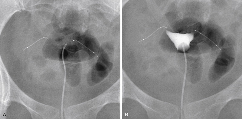
Pictures are taken using a steady beam of X-ray as the dye passes through the uterus and fallopian tubes. The pictures can show problems such as an injury or abnormal structure of the uterus or fallopian tubes, or a blockage that would prevent an egg moving through a fallopian tube to the uterus. A blockage also could prevent the sperms from moving into a fallopian tube and fertilizing an egg.
During HSG, contrast dye outlines the uterine cavity and fallopian tubes. X-ray images help doctors assess the uterine cavity and identify possible blockages or abnormalities.
Uterus and Fallopian Tubes

Contrast Flow Through the Tubes
Evaluation of Tubal and Uterine Anatomy

Review of Uterus and Tubal Findings
The hysterosalpingogram study takes about 15-20 minutes to perform. The test is usually done in the radiology department of a hospital so there is additional time for the woman to register at the facility and fill out a questionnaire and answer questions regarding allergies to medication etc.
The results of the test can be immediately available. The x-ray pictures can usually be reviewed with the patient several minutes after the procedure is done.
Complications commonly associated with a hysterosalpingogram include cramping and spotting.
Rarely HSG may be associated with infections, for which we routinely prescribe prophylactic antibiotics for three days. after the procedure
Find answers to common questions about hysterosalpingography, fallopian tube blockage, procedure, timing, results, side effects and fertility evaluation.
The clinic page states that HSG is best performed immediately after menstruation has completely stopped, typically around Day 7 to Day 9 of the cycle. It should not be performed close to ovulation.
Yes. HSG can show abnormalities in the shape or structure of the uterus and may identify findings such as polyps, fibroids, adhesions, septum or other abnormalities within the uterine cavity.
A normal HSG may show a normally shaped uterus, normal-looking fallopian tubes without obvious scarring or damage, and contrast dye flowing through the tubes and spilling normally into the abdominal cavity.
An abnormal result may show that contrast does not pass through one or both fallopian tubes, suggesting possible blockage, scarring or malformation. It may also show uterine abnormalities such as a septum, polyps or fibroids.
HSG can show certain uterine cavity abnormalities, including growths such as fibroids, particularly when they affect the shape or outline of the uterine cavity.
Yes. Polyps inside the uterine cavity may appear as an abnormality on HSG and may require further evaluation depending on the findings.
HSG may show abnormalities caused by adhesions inside the uterus. Further evaluation may be recommended by the fertility specialist depending on the findings.
HSG is primarily used to evaluate the uterus and fallopian tube patency. The source page lists endometriosis among possible causes associated with blocked fallopian tubes, but HSG itself is not a complete diagnostic test for endometriosis.
Take the first step towards parenthood with compassionate care, expertise, and support tailored to your unique needs.