
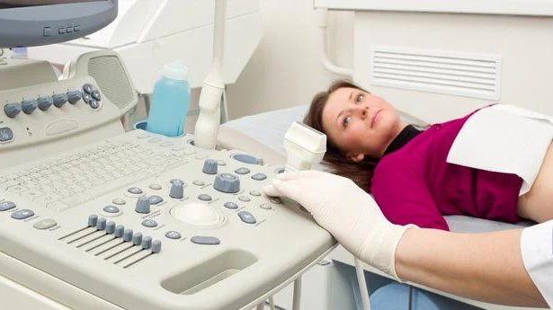
Hysterosonography (HSN)
Hysterosonography, also called sonohysterography, is a non-invasive fertility investigation in which sterile saline solution is slowly introduced into the uterus while ultrasound imaging is performed. The saline gently expands and outlines the uterine cavity, helping the doctor obtain a clearer and more detailed view of the inside of the uterus. It can make structural abnormalities such as polyps, fibroids and adhesions easier to identify than with a regular vaginal ultrasound alone, making it a useful investigation when evaluating uterine factors that may be relevant to fertility and treatment planning.
Key questions
What is Hysterosonography?
Hysterosonography is a non invasive technique that involves the slow infusion of sterile saline solution into a woman’s uterus during ultrasound imaging. The procedure is more likely to reveal structural abnormalities than regular vaginal sonography will show alone.
Hysterosonography allows the doctor to evaluate abnormal growths inside the uterus, abnormalities of the endometrium or disorders affecting deeper tissue layers. Hysterosonography does not require either radiation or contrast media, or invasive surgical procedures
Frequently Asked Questions About Hysterosonography
Find answers to common questions about Hysterosonography (HSN), its purpose, procedure, timing, results and possible side effects.
Hysterosonography, also called sonohysterography, is a non-invasive technique in which sterile saline is slowly infused into the uterus while ultrasound imaging is performed. It helps provide a more detailed view of the uterine cavity than regular vaginal sonography alone.
According to the source, Hysterosonography is done to assess whether fertility problems may be related to polyps, leiomyomas (fibroids) or adhesions inside the uterus.
No. The source specifically states that Hysterosonography does not require radiation. It uses ultrasound imaging instead.
No. The source describes Hysterosonography as a technique that does not require contrast media. Sterile saline is introduced into the uterine cavity to improve visualisation.
According to the source page, Hysterosonography is performed during the first 10 days of the menstrual cycle.
A baseline transvaginal ultrasound is performed first. A sterile speculum is then inserted, the cervix is cleaned and a catheter is placed into the uterine cavity. The speculum is removed, the transvaginal probe is reinserted, and sterile saline is injected through the catheter while ultrasound imaging is performed.
The source states that the ultrasound examination is usually completed within approximately 10 minutes.
Some patients may feel occasional cramping when saline is introduced into the uterus. The source states that pain killers such as Ibuprofen are usually given before the procedure to help prevent cramping.
Yes. Hysterosonography can help identify fibroids or leiomyomas inside the uterus and determine their location, which can be useful when establishing a treatment or management strategy.
Yes. Polyps inside the uterus are among the abnormalities that Hysterosonography can help identify.
Yes. The source specifically lists adhesions inside the uterus as one of the conditions that Hysterosonography can help assess.
A normal result shows that the inside of the uterus appears normal in shape and size, with no polyps, fibroids or other growths. The openings to both fallopian tubes should also look normal.
Abnormal findings may include an abnormal size or shape of the inside of the uterus, scar tissue, uterine polyps, fibroids or other growths, or a closed opening to one or both fallopian tubes.
Yes. A baseline transvaginal ultrasound is performed to view the endometrium, including its thickness, and Hysterosonography provides a more detailed investigation of abnormalities involving the uterine cavity and endometrium.
No. Hysterosonography uses sterile saline and ultrasound to evaluate the uterine cavity, whereas HSG uses radiographic contrast dye and X-ray imaging. The two tests therefore use different techniques and have different purposes.
Ready to begin your journey?
Take the first step towards parenthood with compassionate care, expertise, and support tailored to your unique needs.