The hysteroscopy is an important tool in the study of infertility, recurrent miscarriage, or abnormal uterine bleeding. Diagnostic hysteroscopy is used to examine the inside of the uterus, also known as the uterine cavity, and is helpful in diagnosing abnormal uterine conditions such as internal fibroids, scarring, polyps, and congenital malformations.
What is Hysteroscopy?
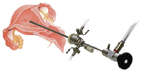
Hysteroscopy is a visual examination of the uterus and it’s lining using a thin viewing Tool called the Hysteroscope which is inserted into the vagina and gently moved through the cervix into the uterus. The hysteroscope has a light and camera hooked to it so your doctor can see the lining of uterus on a video screen.
A hysterosalpingogram (an x-ray of the uterus and fallopian tubes) or an endometrial biopsy may be performed before or after a diagnostic hysteroscopy.
Why is Hysteroscopy Done?
A Diagnostic hysteroscopy may be done to:
- See whether there is a problem in the shape or size of the uterus or if scar tissue in the uterus is causing infertility.
- Find the cause of severe cramping or abnormal bleeding. Your doctor can pass heated tools through the hysteroscope to stop the bleeding.
- Look at the uterine openings to the Fallopian Tubes. If the tubes are blocked, your doctor may be able to open the tubes with special tools passed through the hysteroscope.
- Find the possible cause of repeated miscarriage.
- Find and remove small fibroids or polyps.
- Check for endometrial cancer.
- Use heated tools to remove problem areas in the lining of the uterus.
How is Hysteroscopy Done?
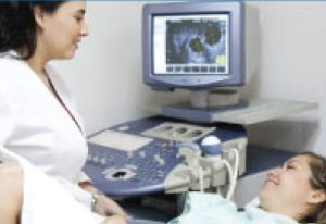
A hysteroscopy is usually done by your gynecologist in the operating room of a hospital or surgery center. Most women are discharged on the same day. Hysteroscopy is usually done on Day 6 – 8 of the cycle after completion of menses.
The procedure is done under local or general anesthesia.
Patient is asked to take off all of their clothes and wear a gown for the test. You will empty your bladder before the test. You will then lie on your back on an examination table with your feet raised and supported by footrests (stirrups).
Your doctor will put an instrument with smooth, curved blades (speculum) into your vagina. The speculum gently spreads apart the vaginal walls so your doctor can see inside the vagina and the cervix. Your vagina will be cleaned with a special soap.
The hysteroscope will be placed at the entrance to the vagina and gently moved through the cervix into the uterus. A gas or liquid will be put through the hysteroscope into your uterus to help doctor see the lining clearly. Then the doctor looks through the hysteroscope at a magnified view of the lining of your uterus. Doctor can also see the uterine openings of the fallopian tubes. A video screen may be used during the test.
If a biopsy or other procedure is done, doctor will use small tools through the hysteroscope. A hysteroscopy takes about 30 minutes, unless other procedures are being done.
What are the risks and complications?
If a fluid is used during the test to help your doctor see the uterine lining clearly, patient may absorb some fluid and feel bloated. It may also change the level of sodium in the patient’s blood. If gas is used, you have a small risk for an air bubble (air embolism) in a blood vessel, though this is very rare.
A hysteroscopy can cause injury to the uterus or cervix, an infection, or bleeding. In rare cases, the uterus, bladder, or bowel can be punctured during the test, requiring surgical repair. If general anesthesia is used, there is a small risk of problems from the anesthesia.
Evaluating the Result
Normal and Abnormal findings are listed below
| Normal: | The inside of the uterus looks normal in shape and size. |
|---|---|
| Absence of polyps, fibroids or other growths in the uterus | |
| Openings to both the Fallopian Tubes look normal. | |
| Abnormal: | The size or shape of the inside of the uterus does not look normal. |
| Presence of Scar tissue in the uterus. | |
| Presence of Uterine polyps, fibroids or other growths. | |
|
The Opening to one or both the fallopian tubes are closed. |