Hysterosonography is a non invasive technique that involves the slow infusion of sterile saline solution into a woman’s uterus during ultrasound imaging. The procedure is more likely to reveal structural abnormalities than regular vaginal sonography will show alone.
Hysterosonography allows the doctor to evaluate abnormal growths inside the uterus, abnormalities of the endometrium or disorders affecting deeper tissue layers. Hysterosonography does not require either radiation or contrast media, or invasive surgical procedures
Why is Hysterosonography Done?
To assess if fertility problems are related to polyps, leiomyomas (fibroids), or adhesions inside the uterus.
How is Hysterosonpgraphy done?
A woman is examined during the first 10 days of the menstrual cycle.
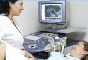
A Baseline transvaginal ultrasound procedure is performed first to view the endometrium, including its thickness and any associated ovarian abnormality.
Sonohysterography is then performed as a more in-depth investigation of the abnormalities and their potential causes. Determining the locations of certain abnormalities, such as fibroids or polyps, can be important when establishing a treatment or management strategy for a patient’s particular condition.
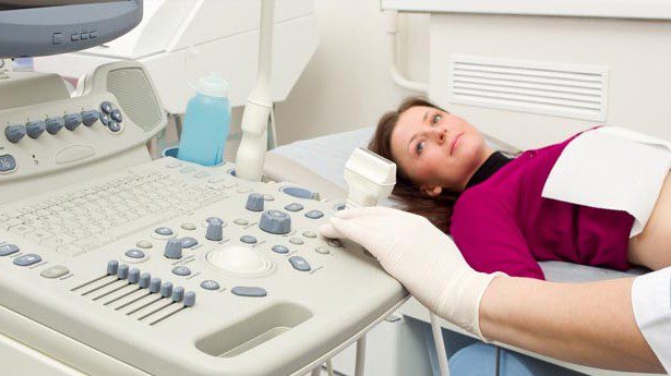
Following the baseline exam, the transvaginal probe will be removed, and a sterile speculum will be inserted as the patient lies on her back with knees bent . The cervix will be cleansed, and a catheter will be inserted into the uterine cavity. Once the catheter is in place, the speculum will be removed, and the transvaginal probe will be re-inserted into the vaginal canal. Sterile saline will then be injected through the catheter into the uterine cavity as ultrasound is being performed.
This ultrasound examination is usually completed within 10 minutes.
What are the risks or Complications?
During the sonohysterogram, patient may feel occasional cramping as a result of the introduction of the saline. Pain Killers like Ibuprofen are usually given before the procedure to prevent cramping. Patient may sometimes have vaginal spotting for a few days after the procedure, which is normal.
Evaluating the Result
Hysterosonography/Sonohysterography
Normal:
The inside of the uterus looks normal in shape and size.
Absence of polyps, fibroids or other growths in the uterus
Openings to both the Fallopian Tubes look normal.
Abnormal:
The size or shape of the inside of the uterus does not look normal.
Presence of Scar tissue in the uterus.
Presence of Uterine polyps, fibroids or other growths.
The Opening to one or both the fallopian tubes are closed.